Corneal Topography
Corneal Topography
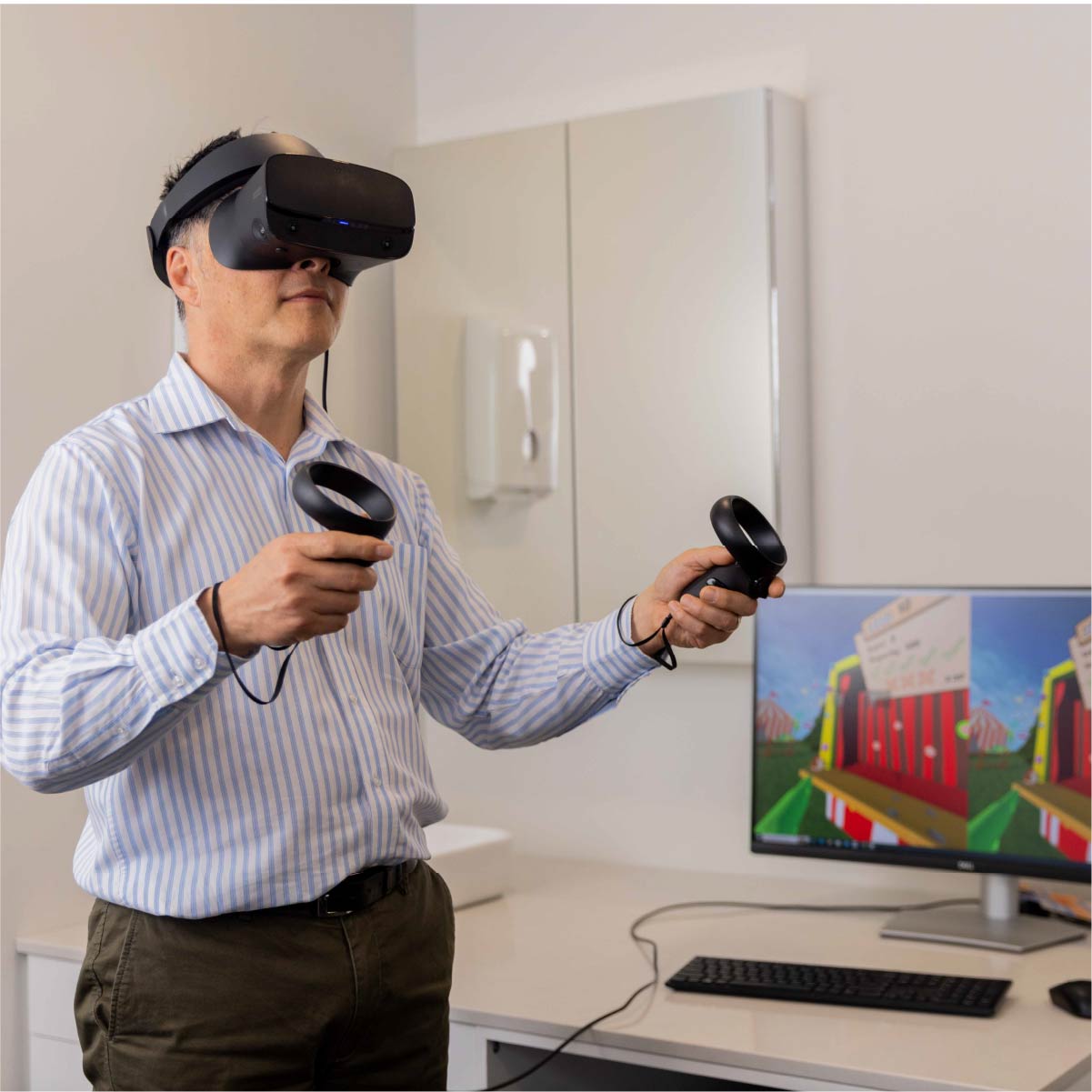
Our Adelaide and Henley Beach practices are both heavily involved in contact lens care and fitting. A crucial aspect of contact lens practice is the precise mapping of your cornea, the transparent front surface of your eye. But why is this mapping so essential, and how do we achieve it?
Exploring Eye Topography
When we talk about topography, we're referring to the process of capturing the shape and unique features of a surface, much like how we map the contours of land (like mountains and valleys.) Similarly, the front surface of your eye, including the cornea, has its own distinct topographical features.
A Trio of Cutting-Edge Technology
To ensure the most accurate and comprehensive assessment of your cornea and the surrounding sclera (the white part of your eye), we've invested in the latest mapping technology. Our practices are equipped with not one, but three distinct pieces of equipment dedicated to this purpose:
- Medmont E300: This advanced device is designed to deliver precise corneal topography, providing critical insights into the shape and structure of your eye's front surface.
- OCULUS Pentacam AXL Wave: This device takes corneal mapping to the next level, offering comprehensive data on the cornea's front and back surfaces, thickness, volume, and more. Its accuracy is unrivalled, making it an invaluable tool for our practice.
- Eaglet Eye: Eye Surface Profiler (ESP): This specialized profiler enhances our ability to assess the eye's surface topography with precision, contributing to a deeper understanding of your eye health.
The Benefits of Advanced Topography
This information is not only essential for fitting contact lenses perfectly but also for monitoring and managing various eye conditions effectively.
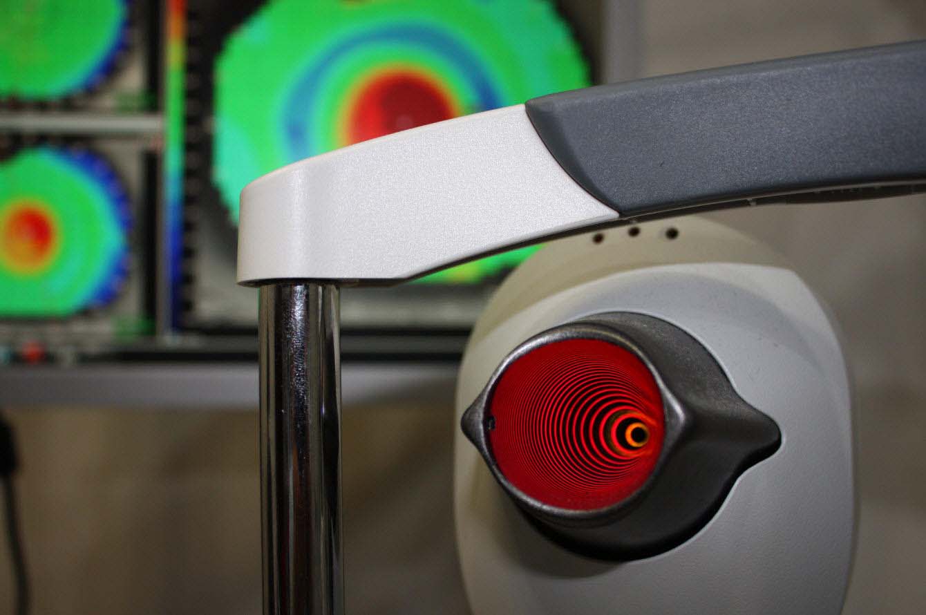
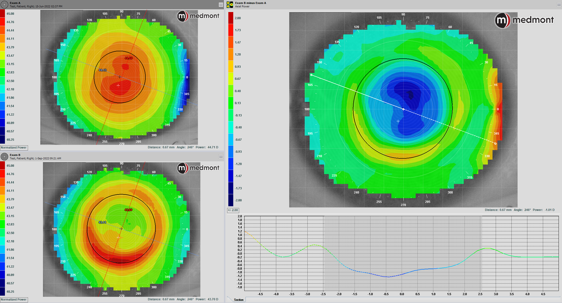
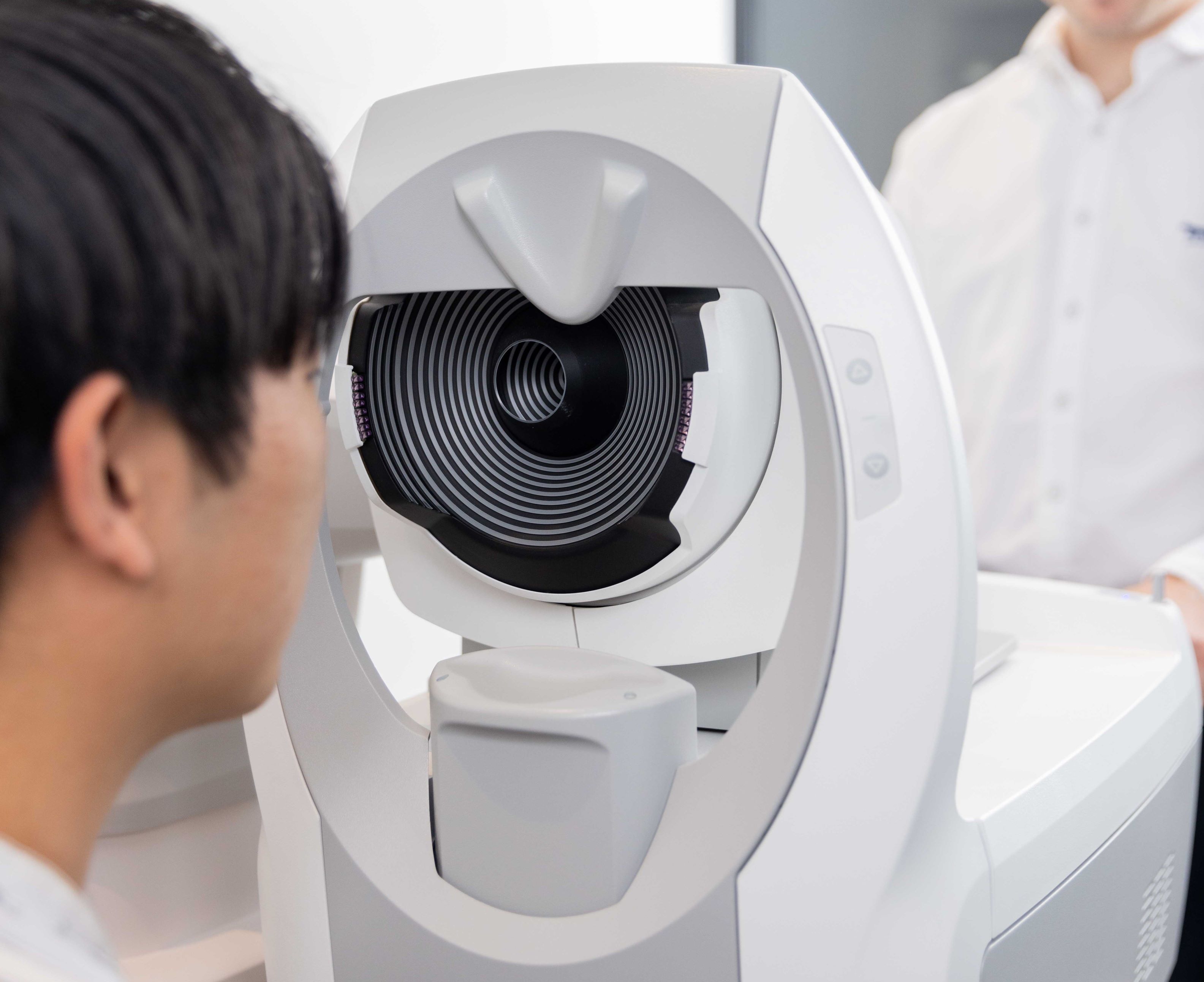
Medmont Corneal Topographer
The Medmont E300 corneal topographer uses Placido disc, or reflection-based, imaging to provide a three-dimensional representation of a patient’s cornea, with particular importance paid to thecornea.1 This information is ideal for fitting rigid gas permeable contact lenses, or RGPs. The most suitable for each patient can be selected – typically through our premier EyeSpace software. This equipment is used extensively for orthokeratology and RGP fitting in keratoconus.

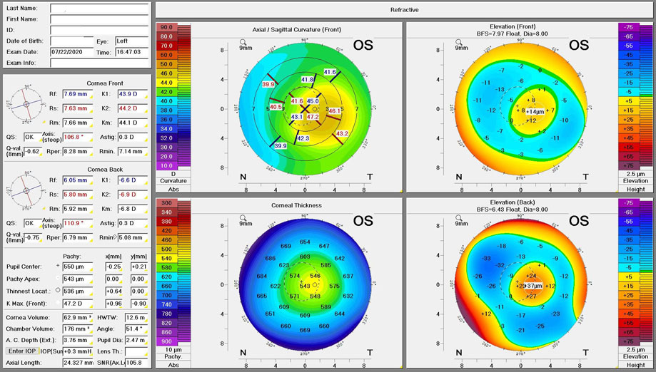
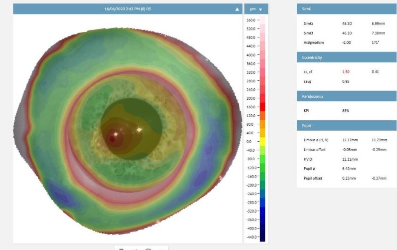
Pentacam AXL Wave
The Pentacam AXL Wave tomographer uses Scheimpflug imaging to map the front and back surfaces of the cornea as well as its thickness and volume. This is a step beyond Placido disc imaging, which maps only the front corneal surface. Its accuracy as a non-contact alternative independent of tear film quality has been proven in countless clinical trials. For this reason, it’s much better for mapping eyes with dry eye disease, and in the design of various custom-made contact lenses including orthokeratology and bespoke RGP lenses. It’s also particularly useful for imaging highly irregular shaped eyes which is common in patients with keratoconus and pellucid corneal marginal degeneration, corneal graft recipients, post corneal cross-linking or in penetrating eye injuries.2, 3, 4
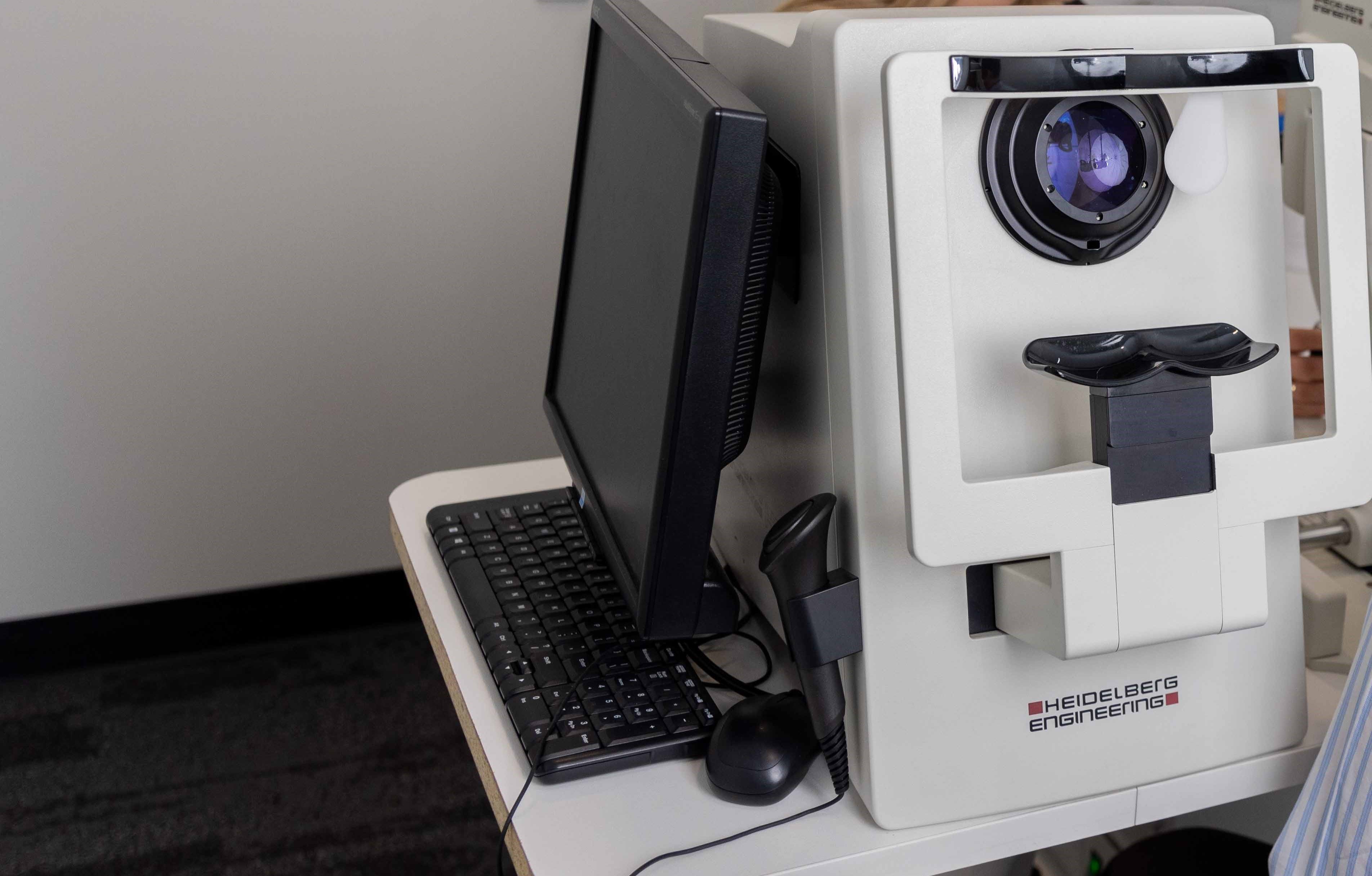
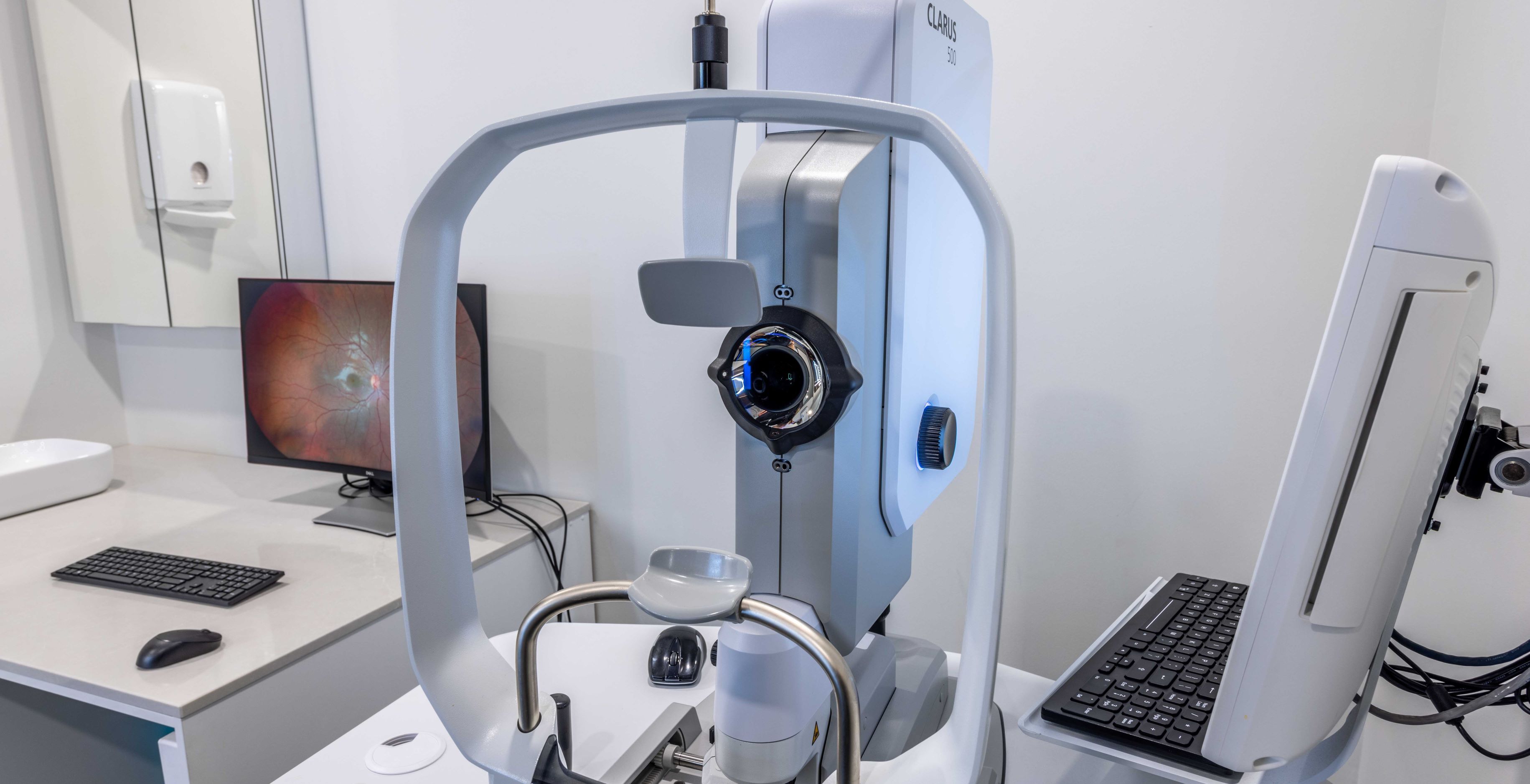
Eaglet Eye ESP
The Eaglet-Eye ESP is a corneo-scleral topographer, meaning it maps out the shape of the front of the eye with incredible accuracy.5, 6 The three-dimensional maps attained by the Eaglet Eye– ESP allows our optometrists to gain a better understanding of how various corneo-scleral disease affects our patient’s eyes and vision.6
The Eaglet-Eye ESP is also very useful for designing custom-made scleral contact lenses.6 These lenses are large diameter lenses that are typically 16-18mm in diameter. Where conventional corneal topographers are limited to an8-10mm diameter scan, the Eaglet-Eye ESP gives us a much bigger picture, with the ability to capture the entire surface of an open eye! 6, 7 This means the Eaglet-Eye ESP will revolutionize the fitting of all contact lenses for anyone with lumps or bumps on their eyes, such as pinguecula or pterygiums.
References
1) Medmont International Pty Ltd. Medmont E300Corneal Topographer User Manual. Australia: Medmont Intl; 2006 Mar:4
2) Van der Worp, E. DeNaeyer, G. Caroline, P.(2017). Understanding Anterior Ocular Surface Shape. Ophthalmology: Current andFuture Developments. 4:68-87.
3) Pinero, D. Martinez-Abad, A. Soto-Negro, R.Ruiz-Fortes, P. Perez-Cambrodi, R. et al. (2019). Differences in corneo-scleraltopographic profile between healthy and keratoconus corneas. Contact lens andAnterior eye. 42(1): 75-84.
4) DeNaeyer, G. Sanders, D. (2018). CollagenCrosslinking for Keratoconus Can Change Scleral Shape. JCLRS. 2(1).
5) Iskander, R. Wachel, P. Simpson, P.Consejo, A. Jesus, D. (2016). Principles of Operation, accuracy and precisionof an Eye Surface Profiler. Ophthalmic and Physiological Optics. 36(3).
6) Iskander, R. (2013). The clinical utilityof the Eye Surface Profiler. IOVS. 54(15).
7) Ritzmann, M. Caroline, P. Borret, R.Korszen, E. (2018). An analysis of anterior scleral shape and its role in thedesign and fitting of scleral contact lenses. Contact Lens and Anterior Eye.41(2): 205-213.
FAQs
Please browse through some of our most frequently asked questions on this topic.


Speak to our friendly team today
Book your appointment now for personalised eye care tailored just for you.